
Relieve Your Back and Neck Pain with San Antonio's Leading Spine Surgeon
Dr. Frank Kuwamura offers personalized, minimally invasive spine treatments to restore your quality of life.
Schedule your appointment today!
Stone Oak
Remington Oaks Medical Building
525 Oak Centre, Suite 140
San Antonio, TX 78258
New innovative treatment option for patients suffering from radical pain due to cervical disc degeneration.
Both cervical fusion and Mobi-C Cervical Disc replace the damaged disc(s) and try to match the healthy disc height, removing pressure on the nerves. Fusion surgery typically requires an anterior metal plate to keep the interbody spacer in place and to stop movement at the surgical level(s).
In contrast, Mobi-C fits entirely within the disc space and is designed to maintain neck movement, which may help reduce adjacent levels from degenerating.
What Is Sciatica?
Our Medical Specialties
ORTHOPEDIC SPINE RECONSTRUCTIVE SURGERY
MINIMALLY INVASIVE ORTHOPEDIC SPINE SURGERY
ORTHOPEDIC SURGERY
CERVICAL SPINE MYELOPATHY
Procedures

ANTERIOR CERVICAL DISCECTOMY & FUSION
What is an ACDF procedure?
An Anterior Cervical Discectomy and Fusion (ACDF) procedure is a type of cervical spine surgery from the front (anterior) of the neck (cervical) that often successfully addresses spinal symptoms. ACDF surgery is a very common procedure relative to overall spine surgeries and has a long and studied record of positive outcomes.
An ACDF surgery consists of removing the damaged disc and then growing bone between the vertebrae above and below. ACDF procedures may be performed with the use of an implant, such as a plate, to provide support until fusion occurs.

EXTREME LATERAL INTERBODY FUSION
What is an eXtreme Lateral Interbody Fusion (XLIF®) procedure?
The eXtreme Lateral Interbody Fusion (XLIF) technique is a minimally disruptive surgical procedure performed through the side of the body. It is designed to treat a range of spinal pathologies. Using patented nerve monitoring technology, the surgeon gains lateral (side) access to the spinal column, avoiding any major nerves in the area between the incision and the column.
The XLIF procedure does not require an anterior (front) or posterior (back) exposure, and thereby does not present the same risks of vascular and/or neural injury as traditional approaches.

ANTERIOR LUMBAR INTERBODY FUSION
What is an Anterior Lumbar Interbody Fusion (ALIF) procedure?
ALIF is a procedure used to treat problems such as disc degeneration, spine instability, and deformities in the curve of the spine. In this procedure, the surgeon works on the spine from the front (anterior) and removes a spinal disc in the lower (lumbar) spine. The surgeon inserts a bone graft into the space between the two vertebrae where the disc was removed (the interbody space).
The goal of the procedure is to stimulate the vertebrae to grow together into one solid bone, a process known as fusion. Fusion creates a rigid and immovable column of bone in the problem section of the spine. This type of procedure attempts to reduce back pain and other symptoms

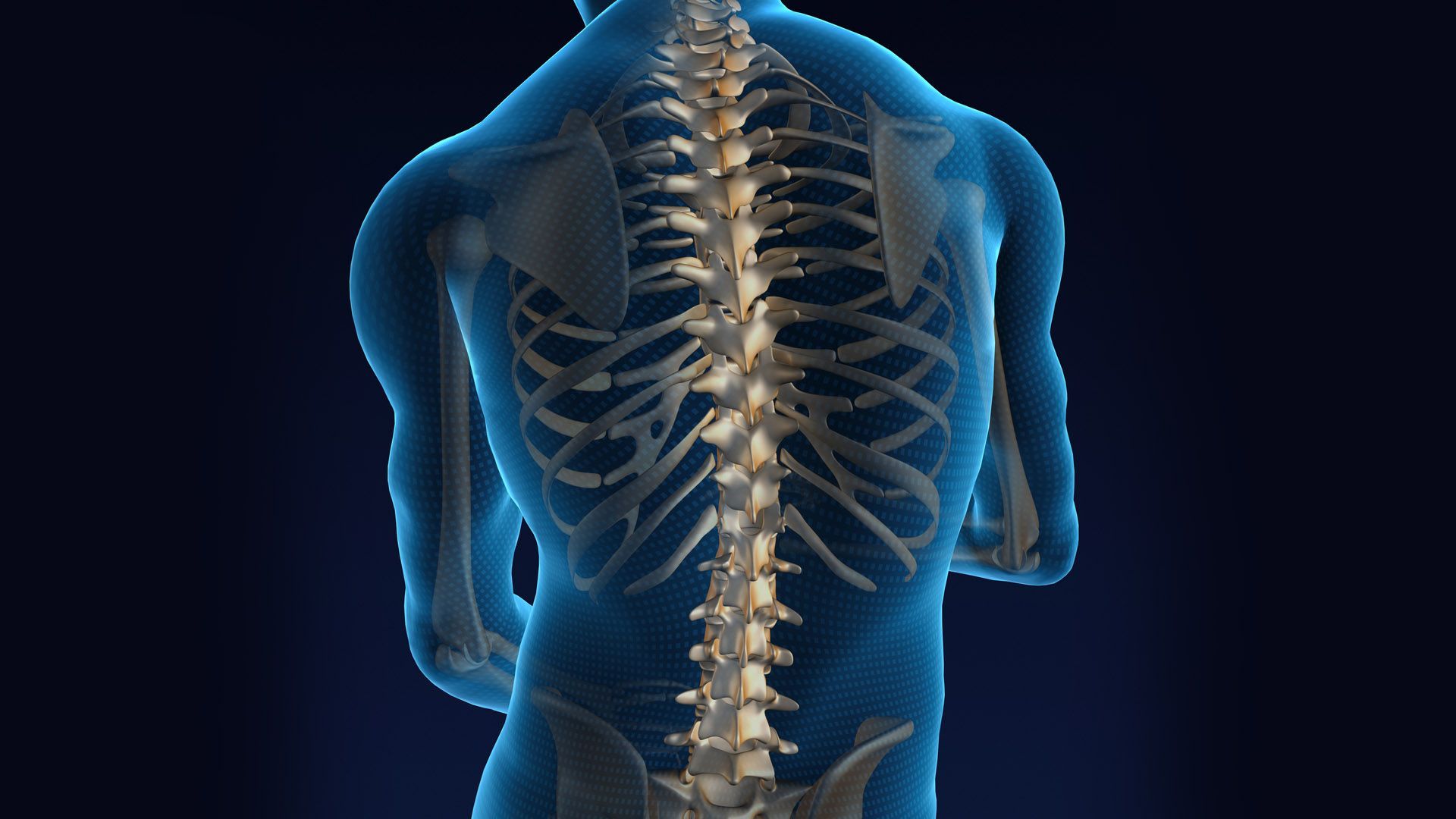
The Healthy Neck
The Mobi-C Cervical Disc:
The neck (cervical spine) is made up of the bones (vertebrae), spinal cord, nerves, muscles, ligaments, and the system that carries blood (blood vessels). The top seven vertebrae make up the cervical spine and begin at the base of the skull. The vertebrae of the cervical spine protect the spinal cord and support the skull. A disc between each vertebra helps to cushion the vertebrae from moving together with the load of the body. Each disc has a strong outer ring (annulus fibrosus). The outer ring helps keep the disc’s soft center (nucleus pulposus) in place. Disc problems can start from over-use, an accident, or just the wear and tear of everyday life.
The vertebrae and the discs allow a healthy cervical spine to:
- Bend side-to-side (lateral bend).
- Bend forward-to-back (flexion and extension).
- Turn left-to-right (rotation).

Lumbar Stenosis Utilizing METRx® System
Decompression
In preparation for a spinal stenosis procedure, insert the appropriate-sized METRx® System Tube as previously described. Once the Tube is positioned, it is important to extend the laminotomy cephalad above the insertion of the ligamentum flavum.
This is to ensure resection of all hypertrophied ligamentum. After the cephalad border of the ligamentum is exposed, it can be separated from the dura using Angled Curettes or a right or left Ball Tip Dissector. The ligamentum is then resected with a 90° or 40° Kerrison Punch. To ensure complete decompression of the lateral recess, the lateral exposure should allow palpation of the inferior pedicle.

IFUSE IMPLANT SYSTEM
Do you have SI Joint Problems?
The SI joint is a significant cause of low back pain. Clinical publications have identified the SI joint as a pain generator in 15-30% of chronic low back pain patients (*1-4). In addition, the SI joint is a pain generator in up to 43% of patients with continued or new onset low back pain after a lumbar fusion (*5).
Like any other joint in the body, the SI joint can be injured and/or become degenerative. When this happens, people can feel pain in their buttock and sometimes in the low back and legs. This is especially true while lifting, running, walking or even sleeping on the involved side.
Our Specialist
Frank K. Kuwamura, III, M.D.
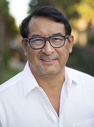
A leader and instructor in emerging technology and spine surgery, Dr. Kuwamura treats multiple conditions of the cervical, thoracic, and lumbar areas of the spine, including: fractures, spinal stenosis, arthritis, tumors, spinal deformities, sciatica, spinal infections, and much more. Dr. Kuwamura is fellowship trained in spine treatments from the Florida Neck and Back Institute and has been in practice for over 19 years and maintains his board certification through ongoing education and training.
Beginning his career as an orthopedic surgeon, Dr. Kuwamura is a graduate of the Boston University School of Medicine and received a Master’s Degree in Microbiology. During his distinguished career, he also served at the National Naval Medical Center in Bethesda, Maryland. Dr. Kuwamura is a pioneer and product designer for leading medical technology and is on-site at MD Spine Care to provide one-on-one consultations and patient-centered care. He is Board Certified with the American Academy of Orthopedic Surgeons and Bexar County Medical Society.
Leave Dr. Kuwamura a review on Google by clicking the button below:
Reviews

Get a customized spine treatment plan.
Office Hours
MONDAY – FRIDAY
8 am – 5 pm
For Billing questions or concerns please contact:
